
E/I balance
the most accurate and largest reconstruction yet
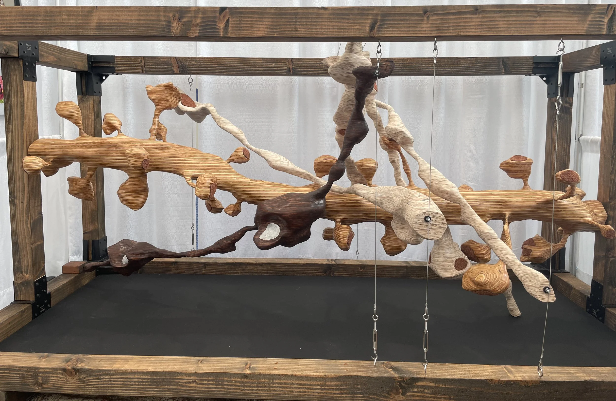
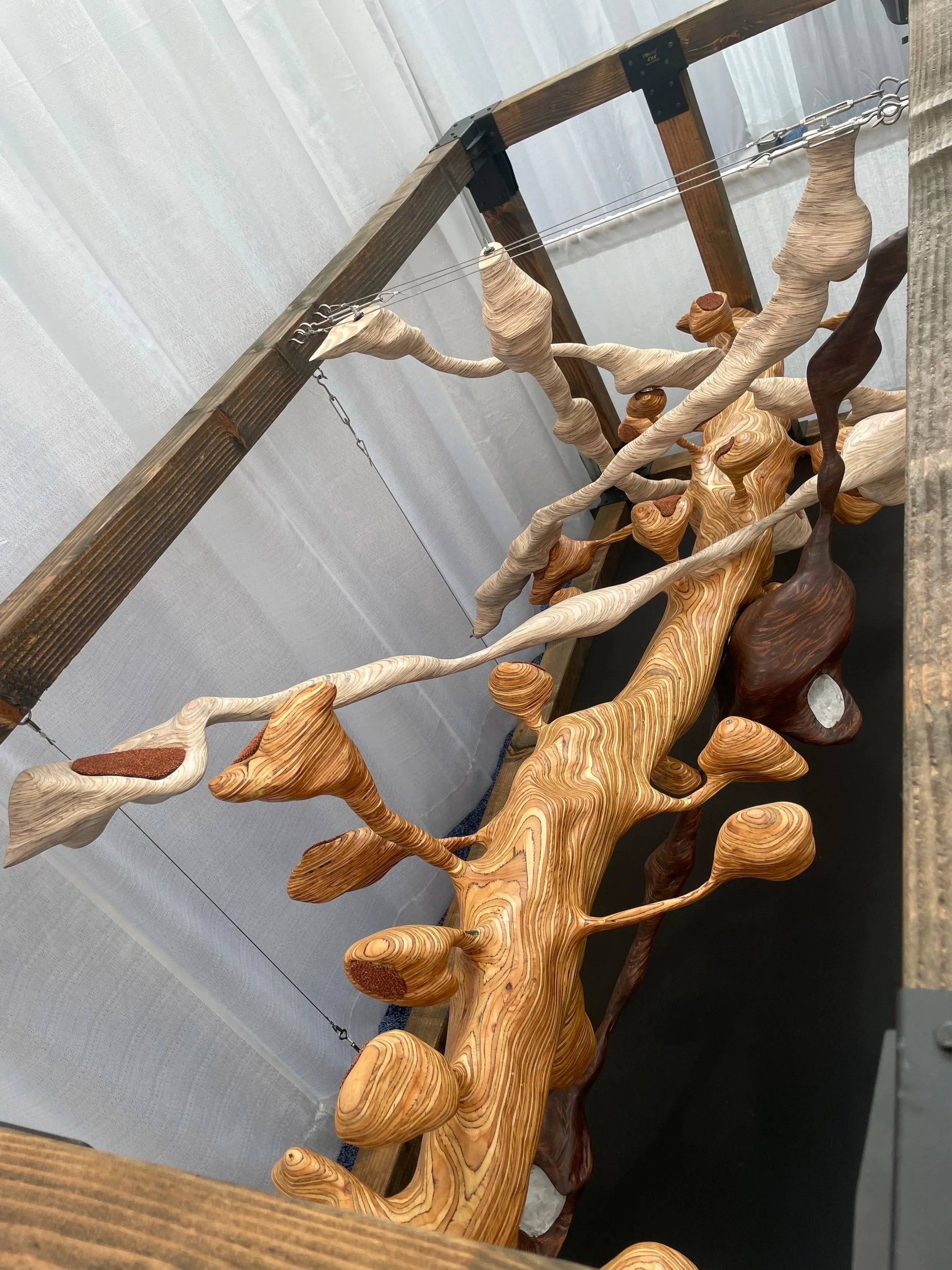
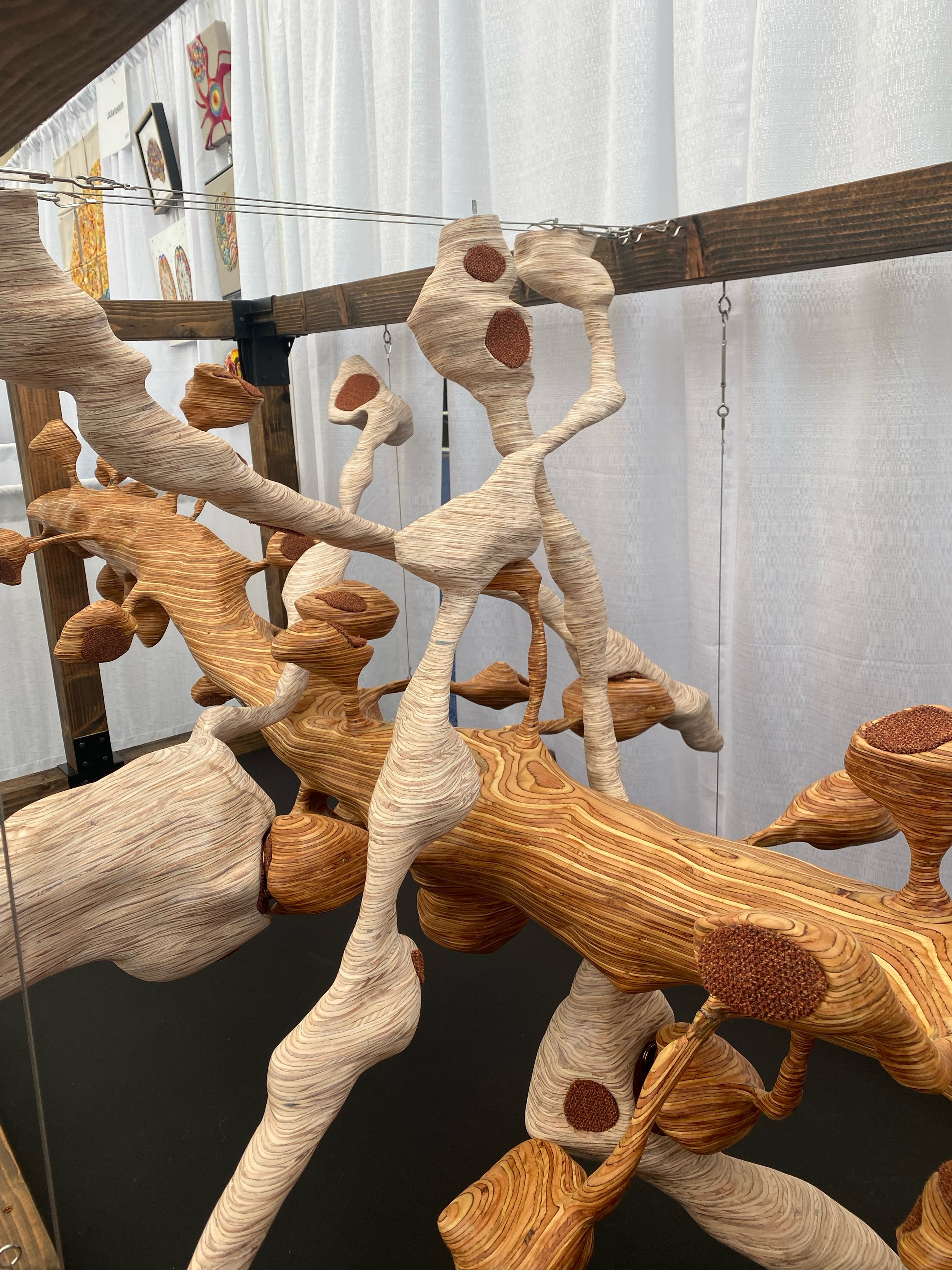
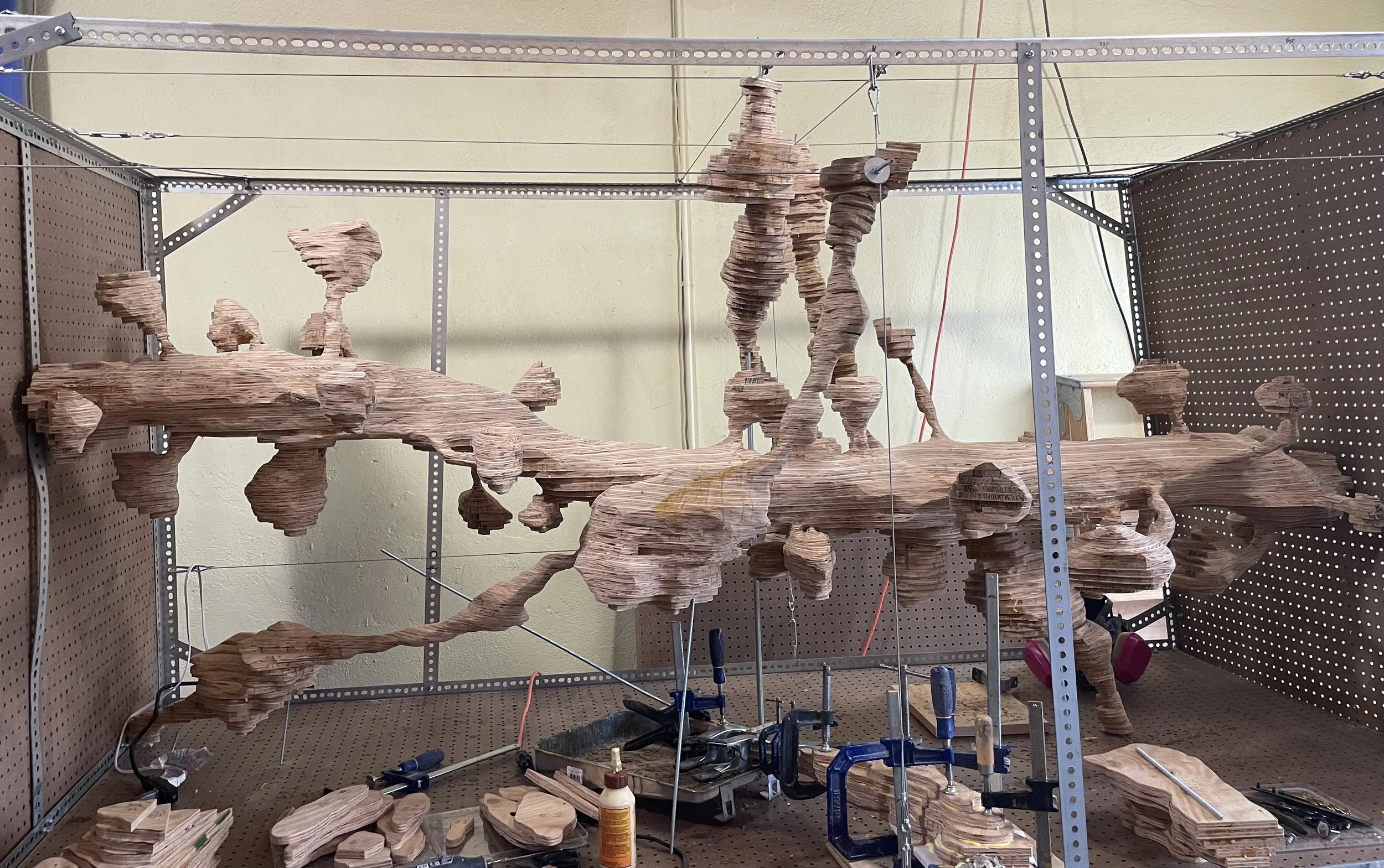
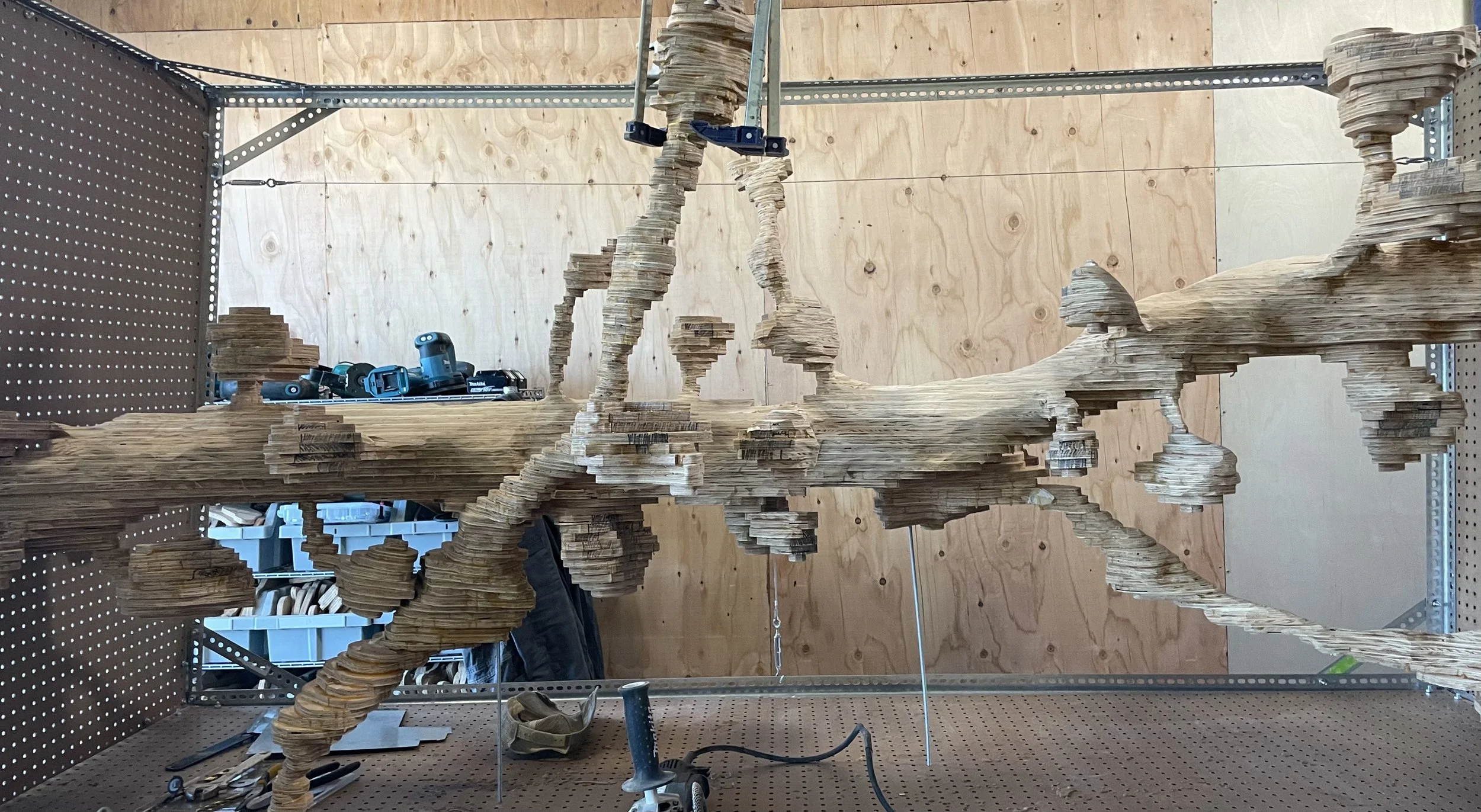
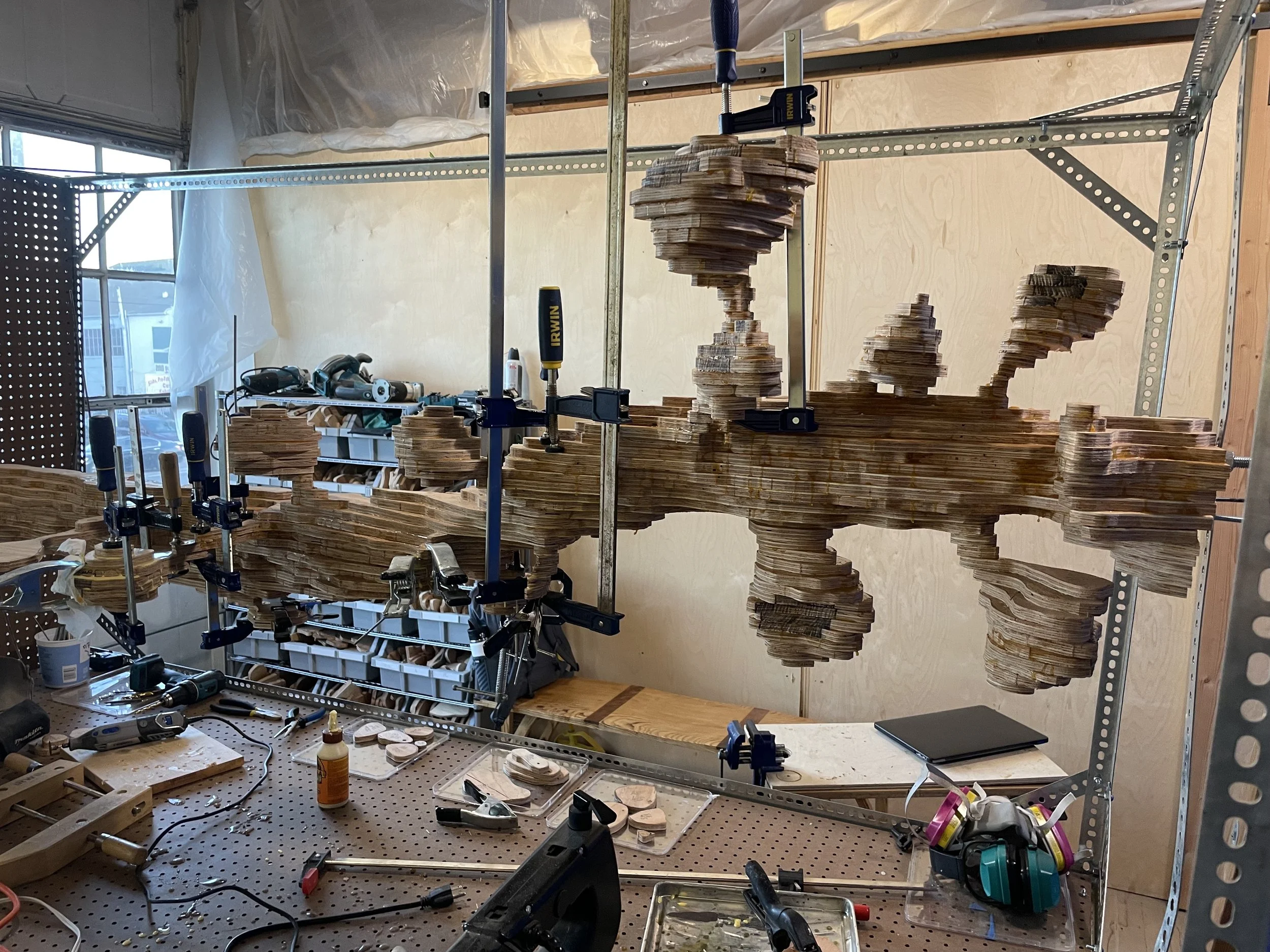
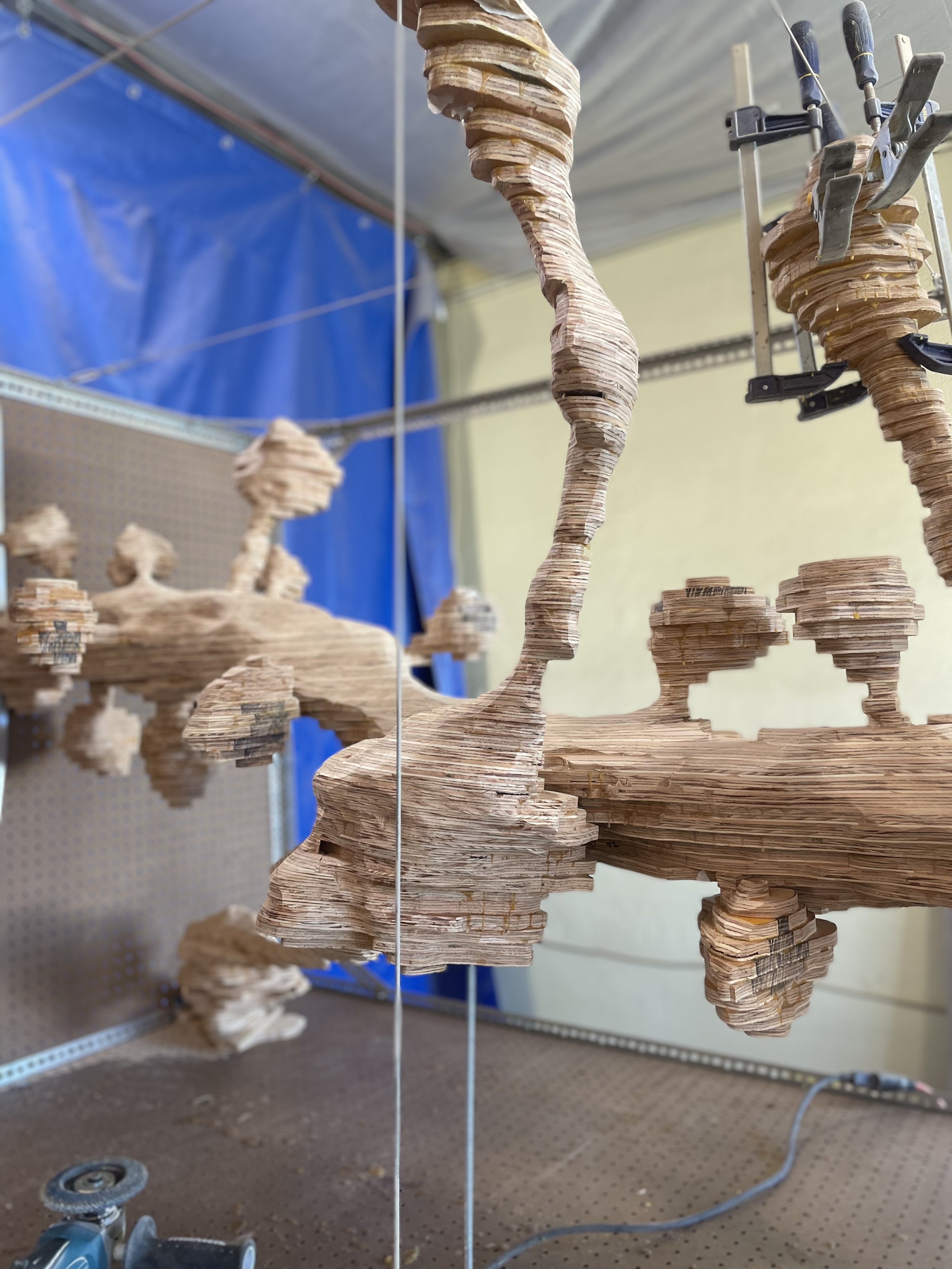
This piece represents a small stretch of dendrite receiving synaptic innervation by excitatory and inhibitory axons in the stratum oriens of the adult mouse hippocampus CA1. The position of each synaptic contact is represented using steel and fabric along the lighter excitatory axons. The darker axon represents a dendritic-targeting inhibitory axon. I’ve been studying the distribution of these synaptic contacts in the lab and wanted to develop a deeper intuitive sense of the spatial relationships between them.
On display at SfN2025 in San Diego

Excitatory spine contacts
On display at SfN2025
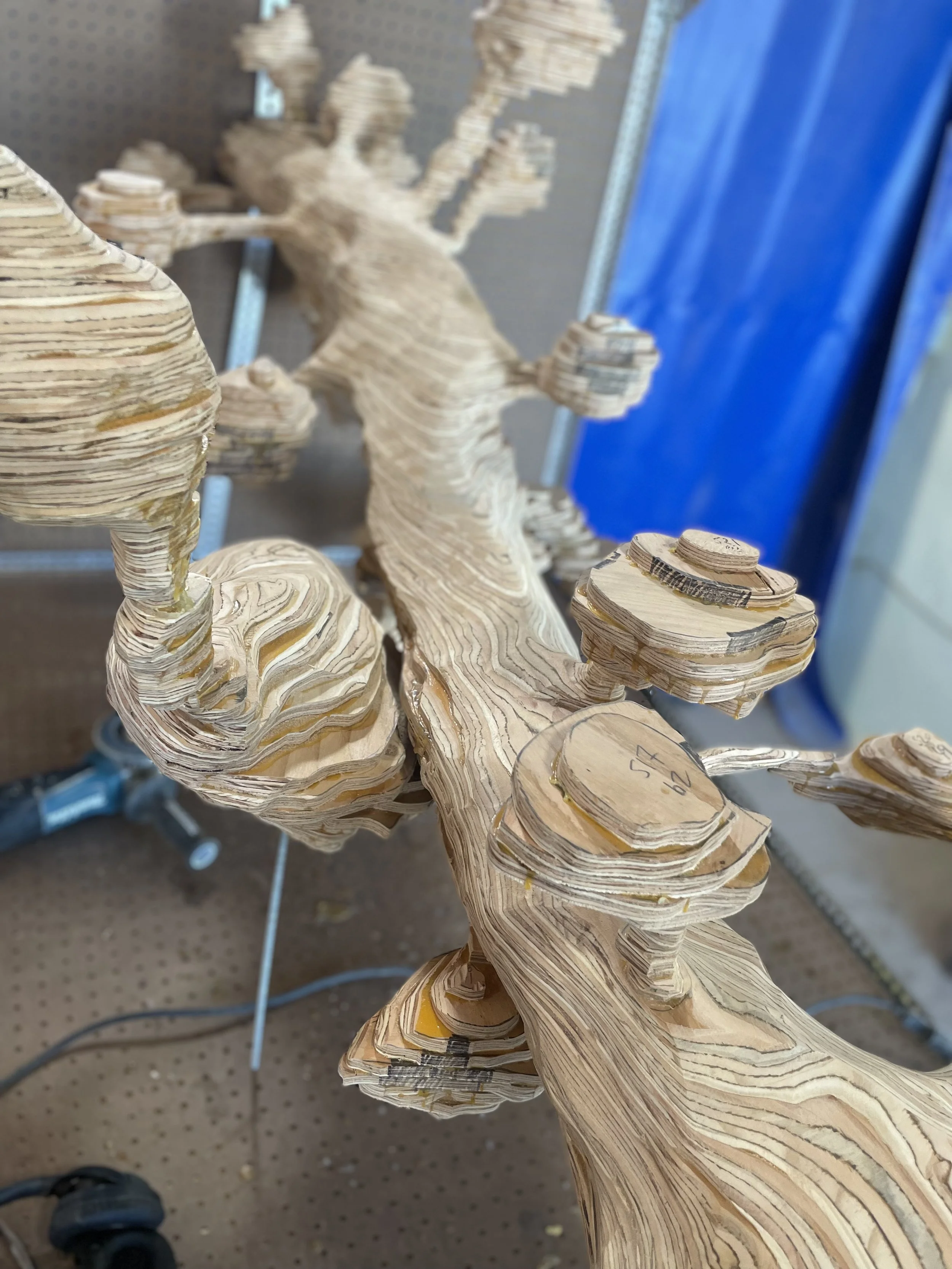
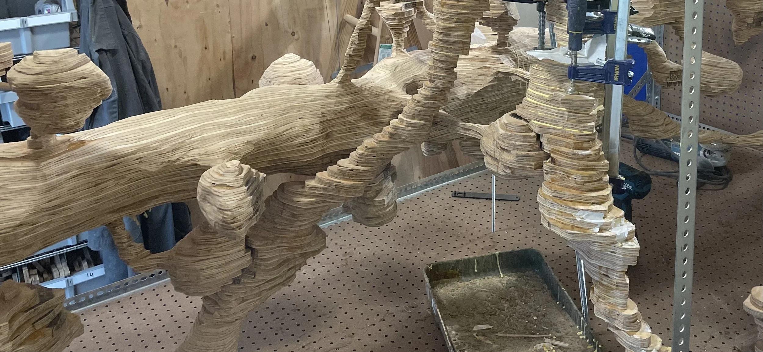
Looking more like membranes and less like lego.
Polishing the GABAergic bouton
Excitatory axon under construction
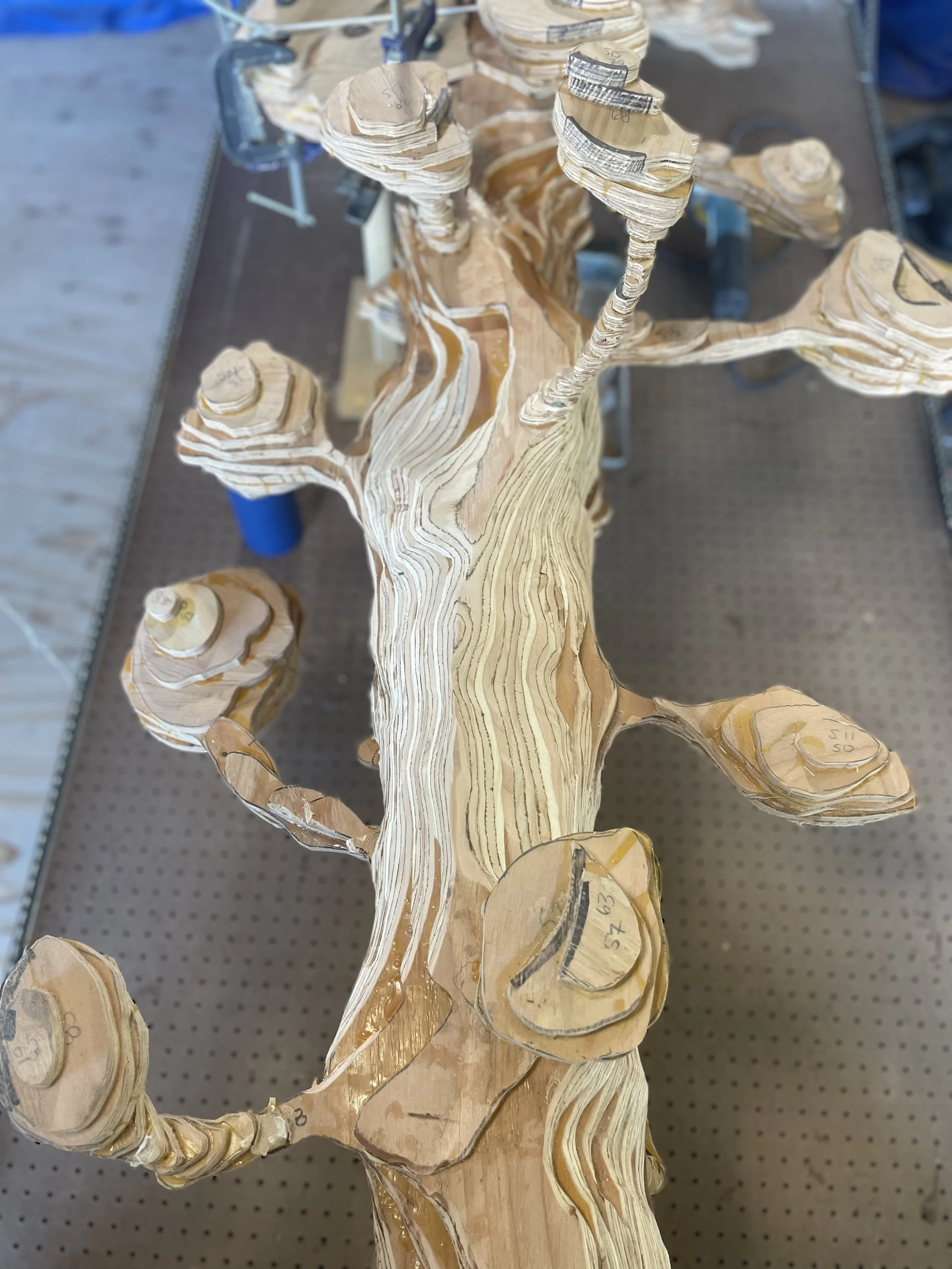
Spines under construction
Building the inhibitory synapse
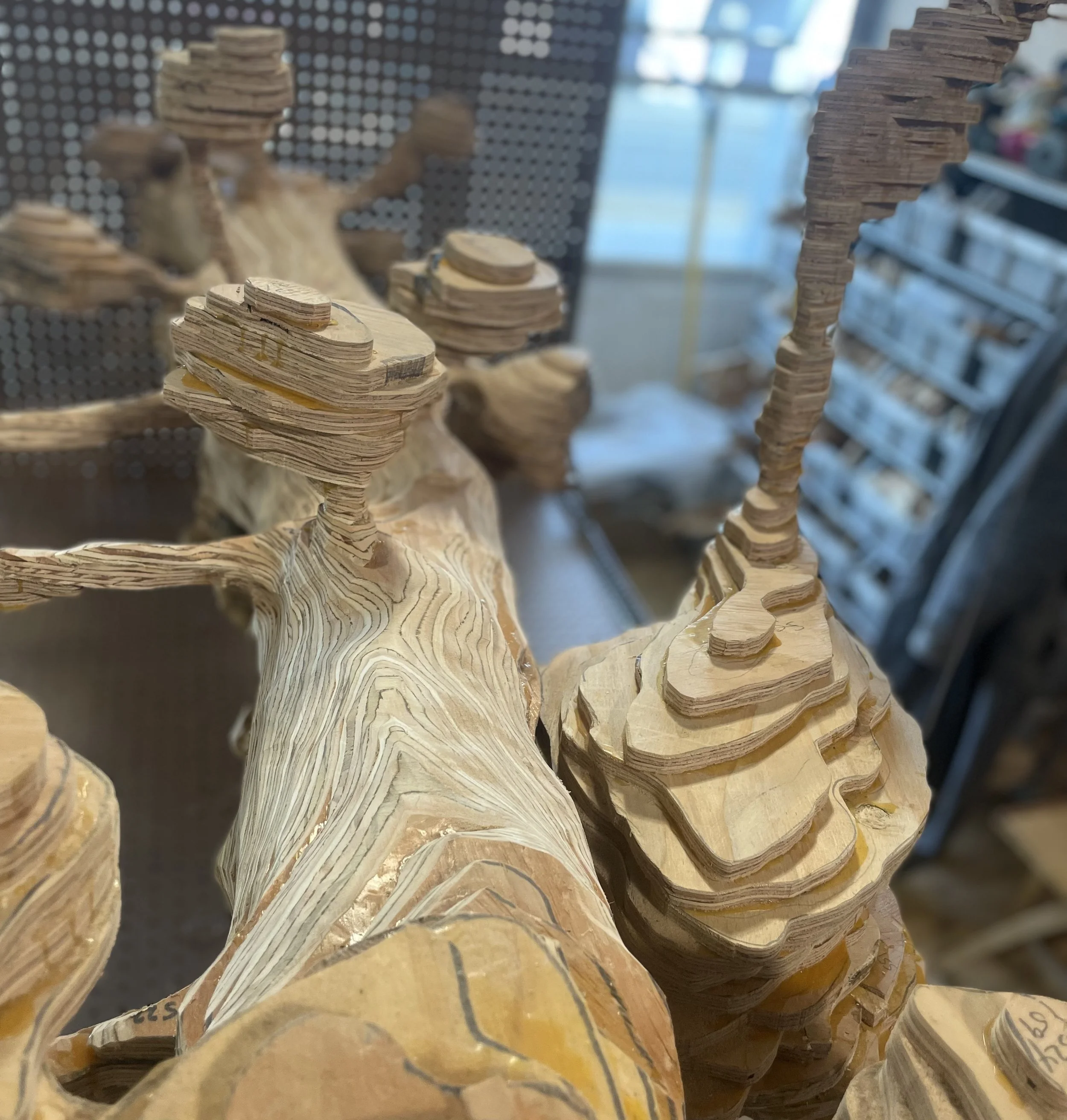
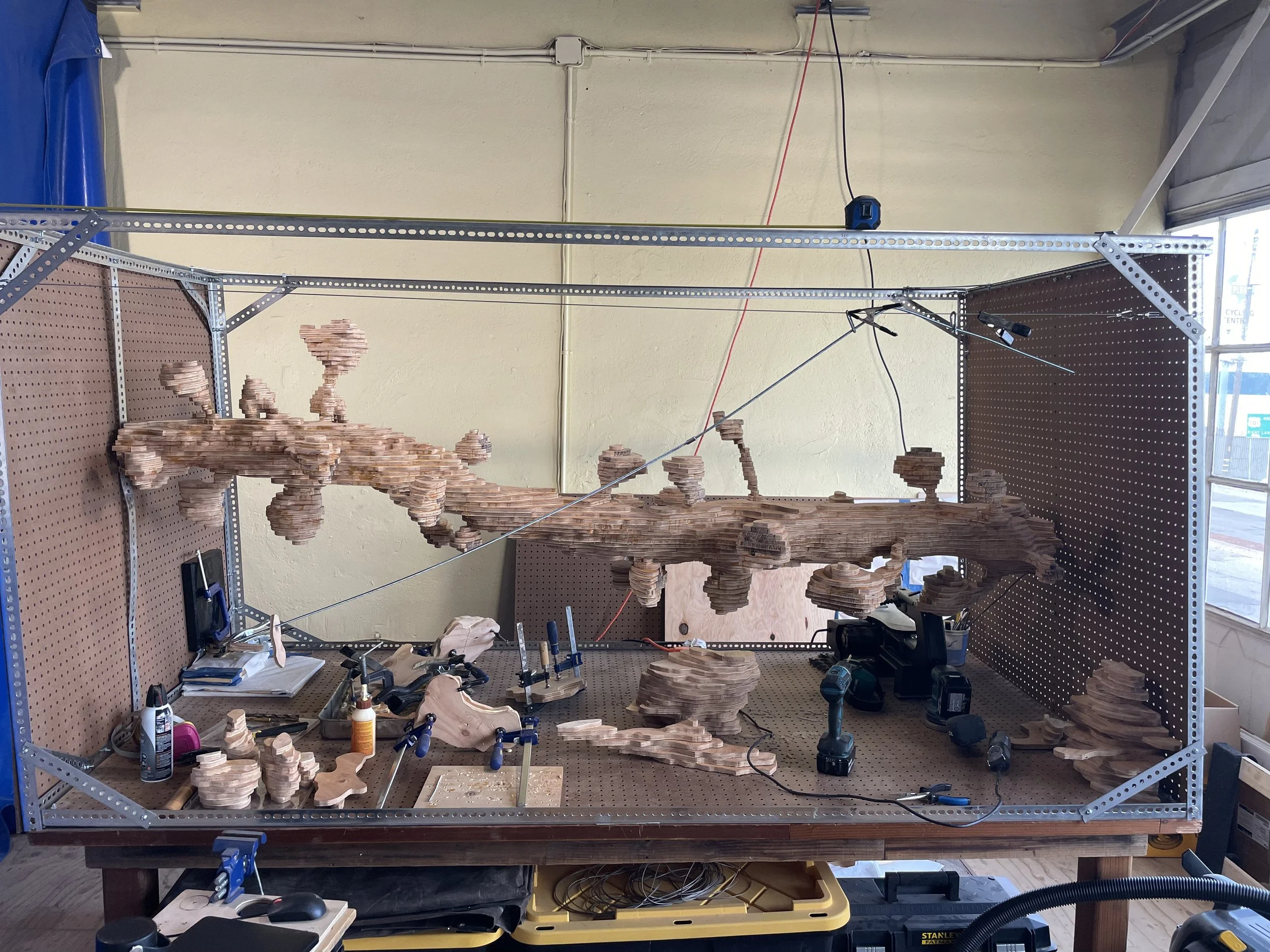
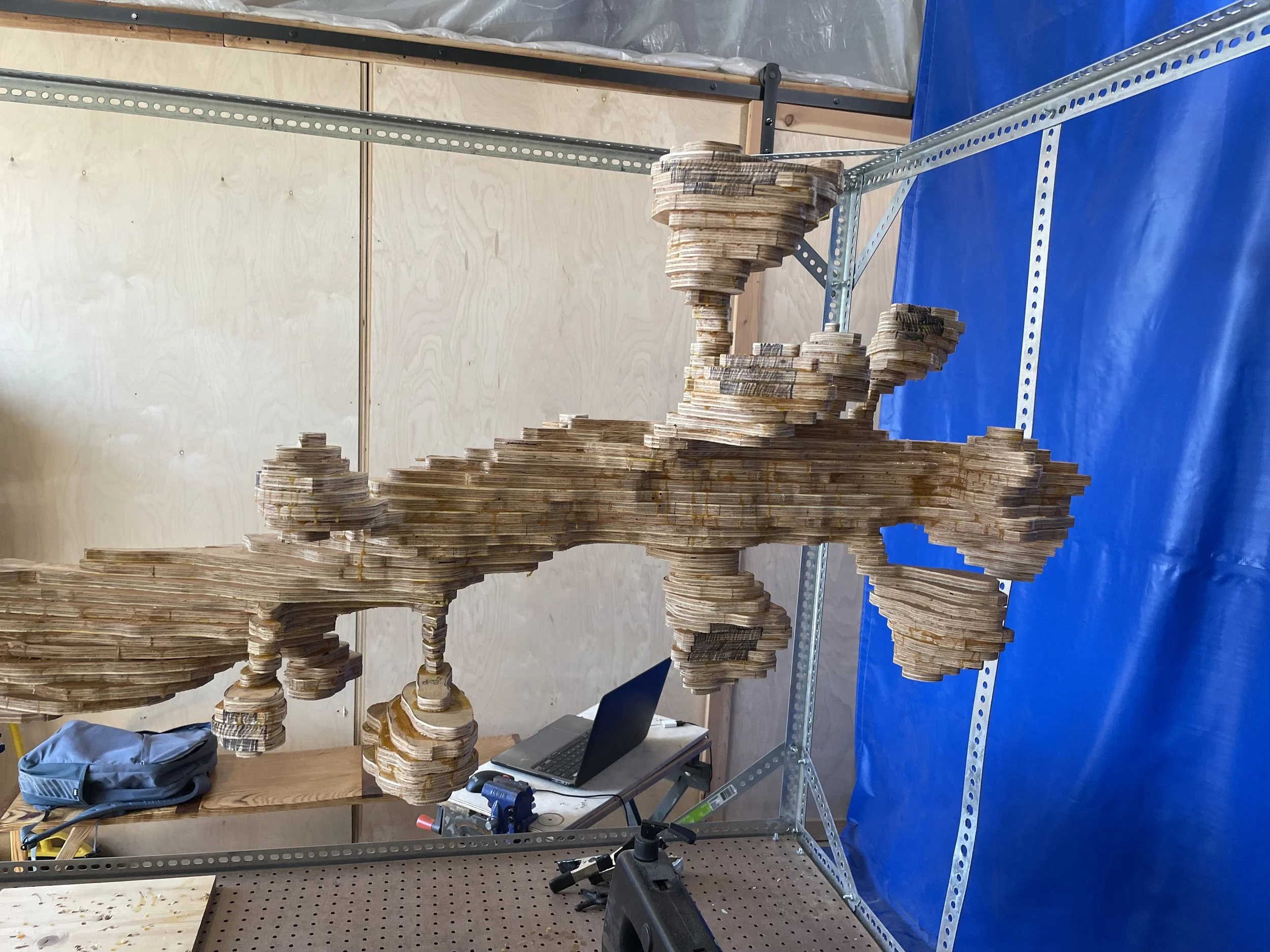
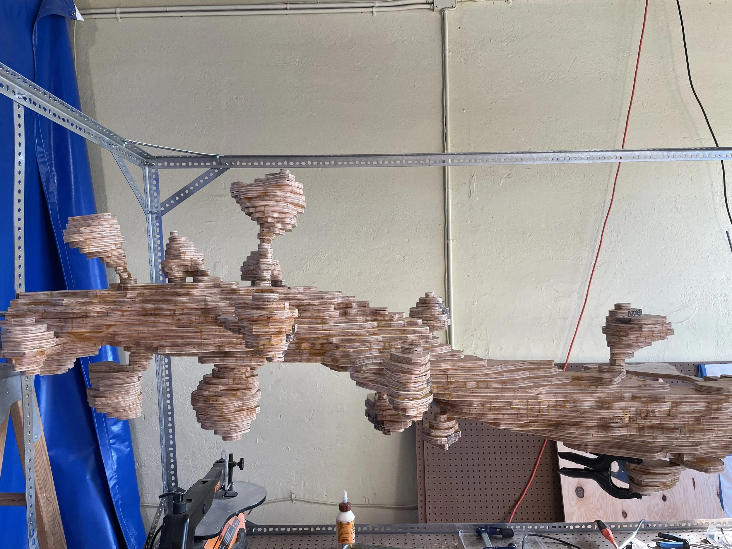
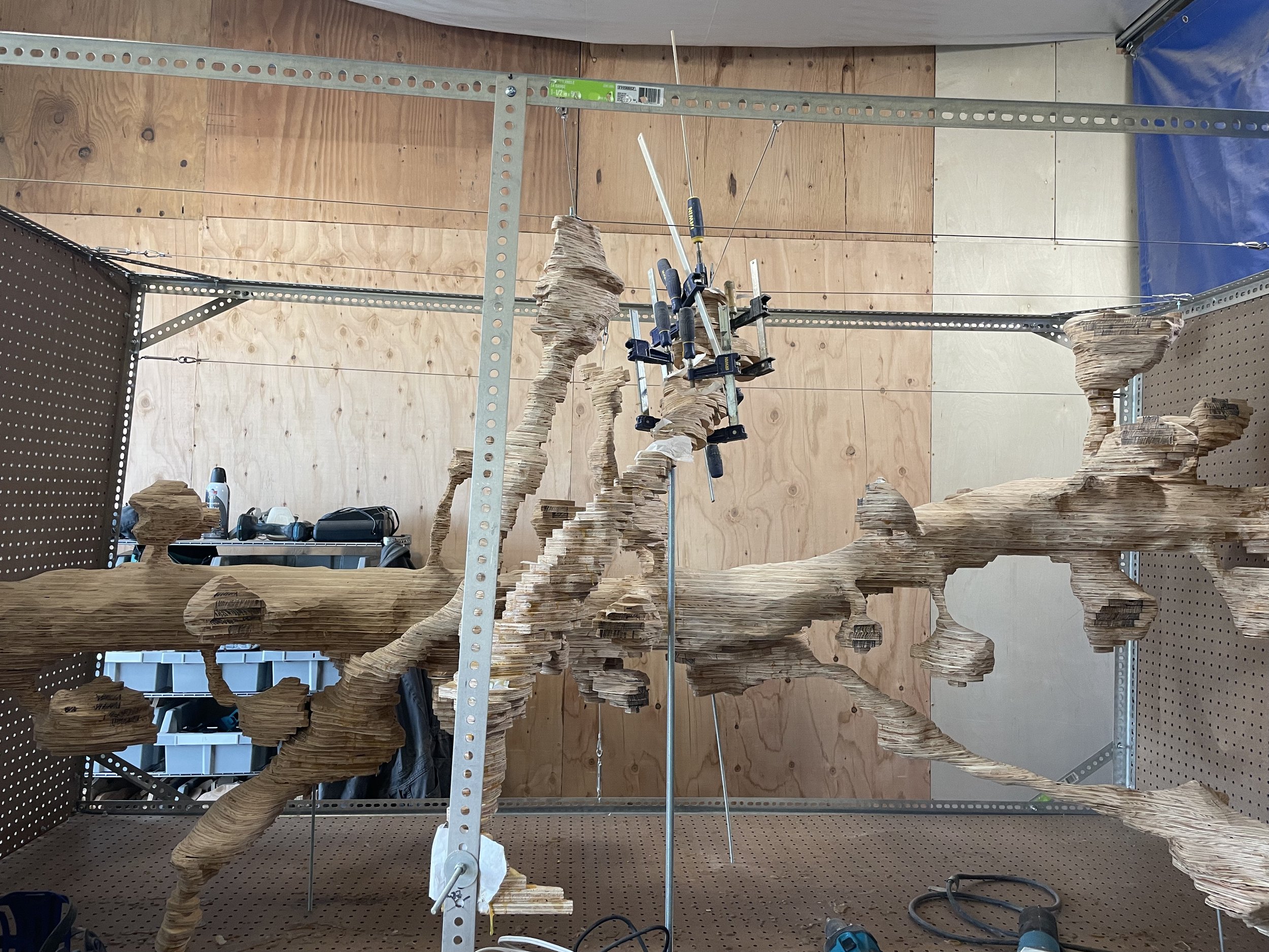
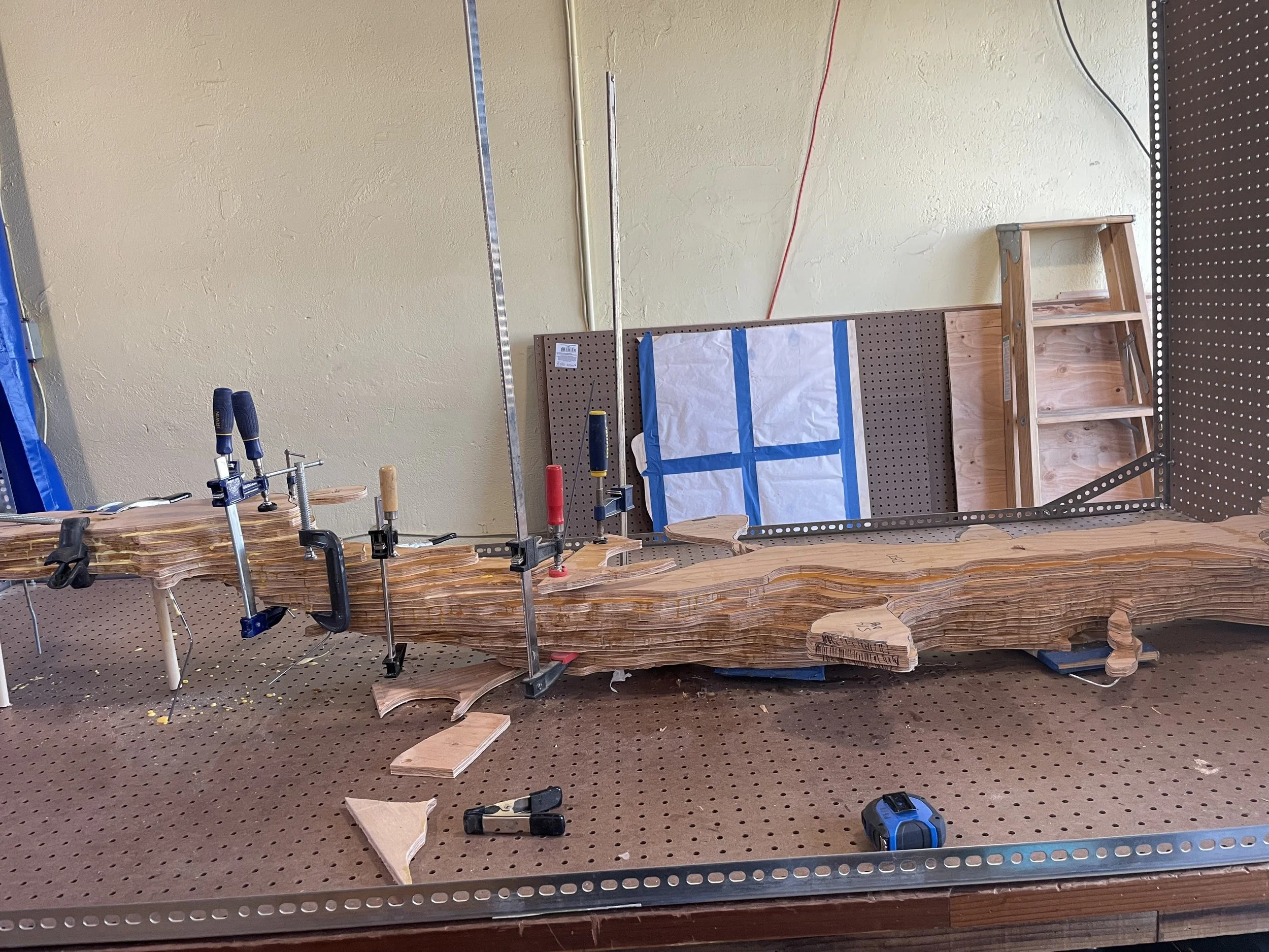
Layered plywood creates a synaptic topography
Building the inhibitory axon
Building an excitatory spine synapse
Synaptic topography
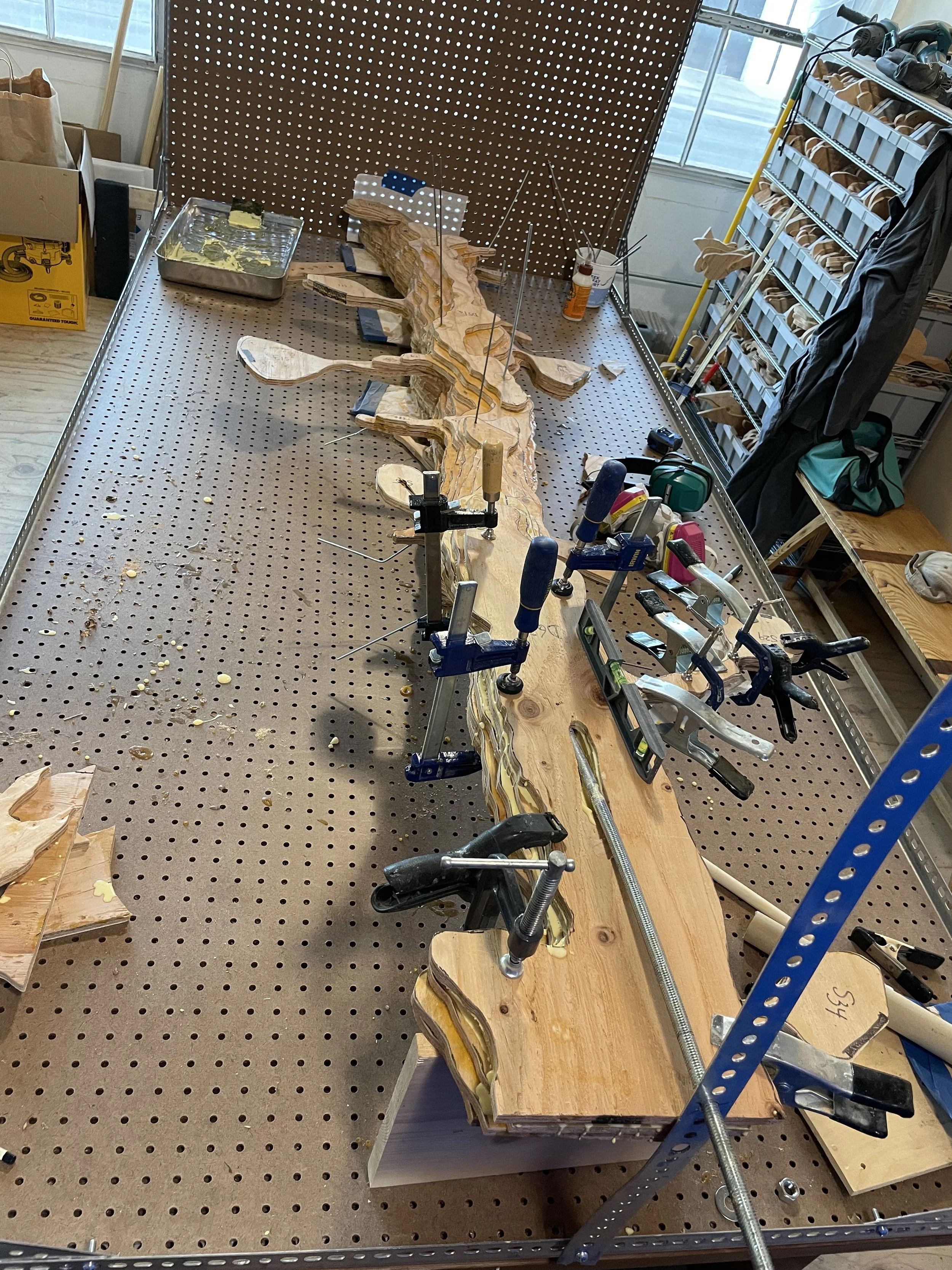
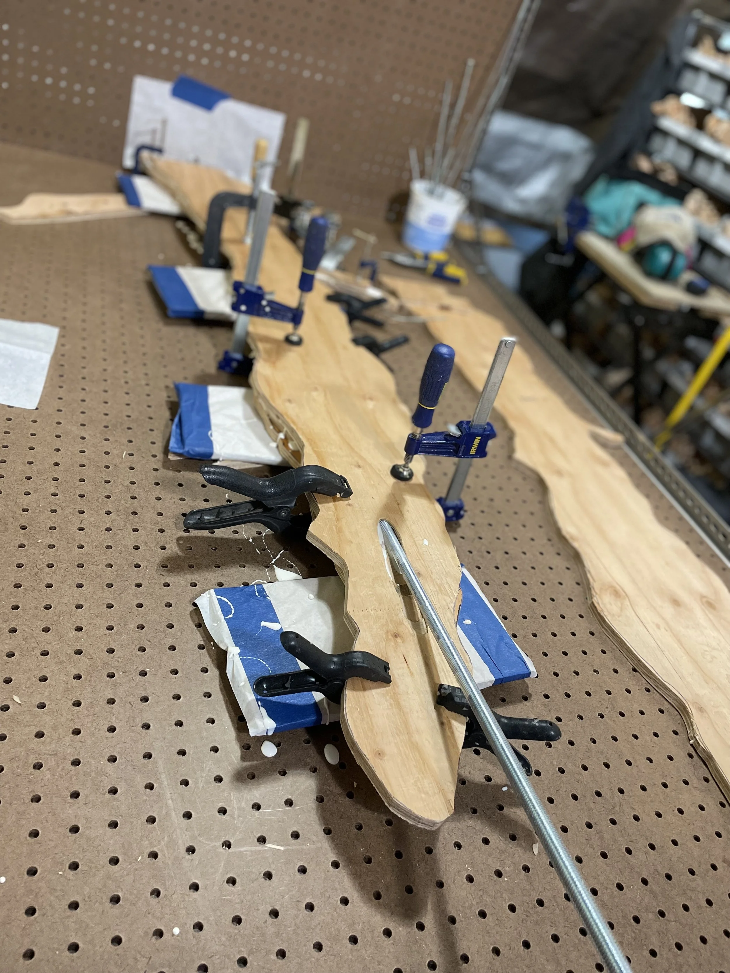
Slowing laying the spines in along the dendrite
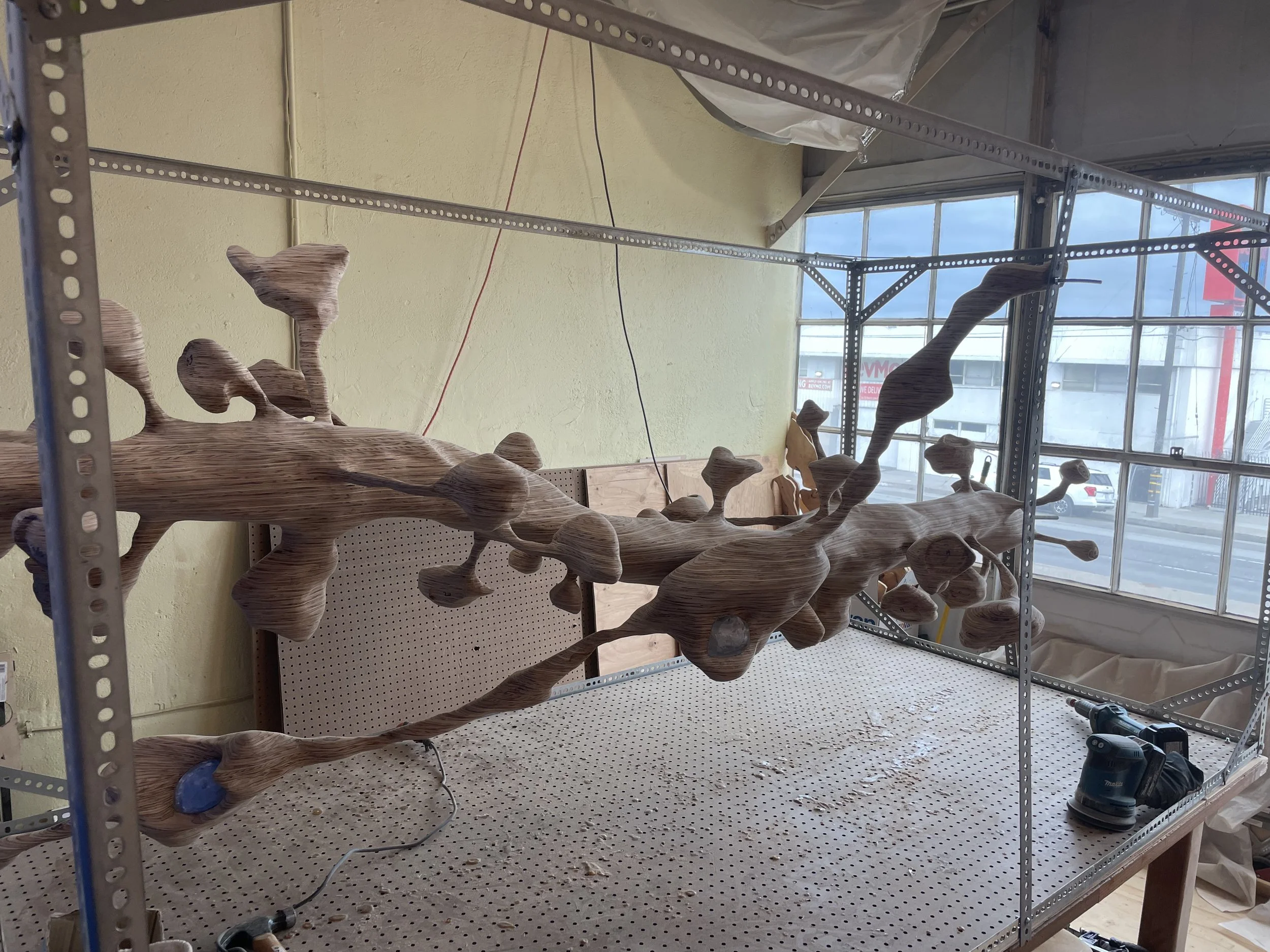
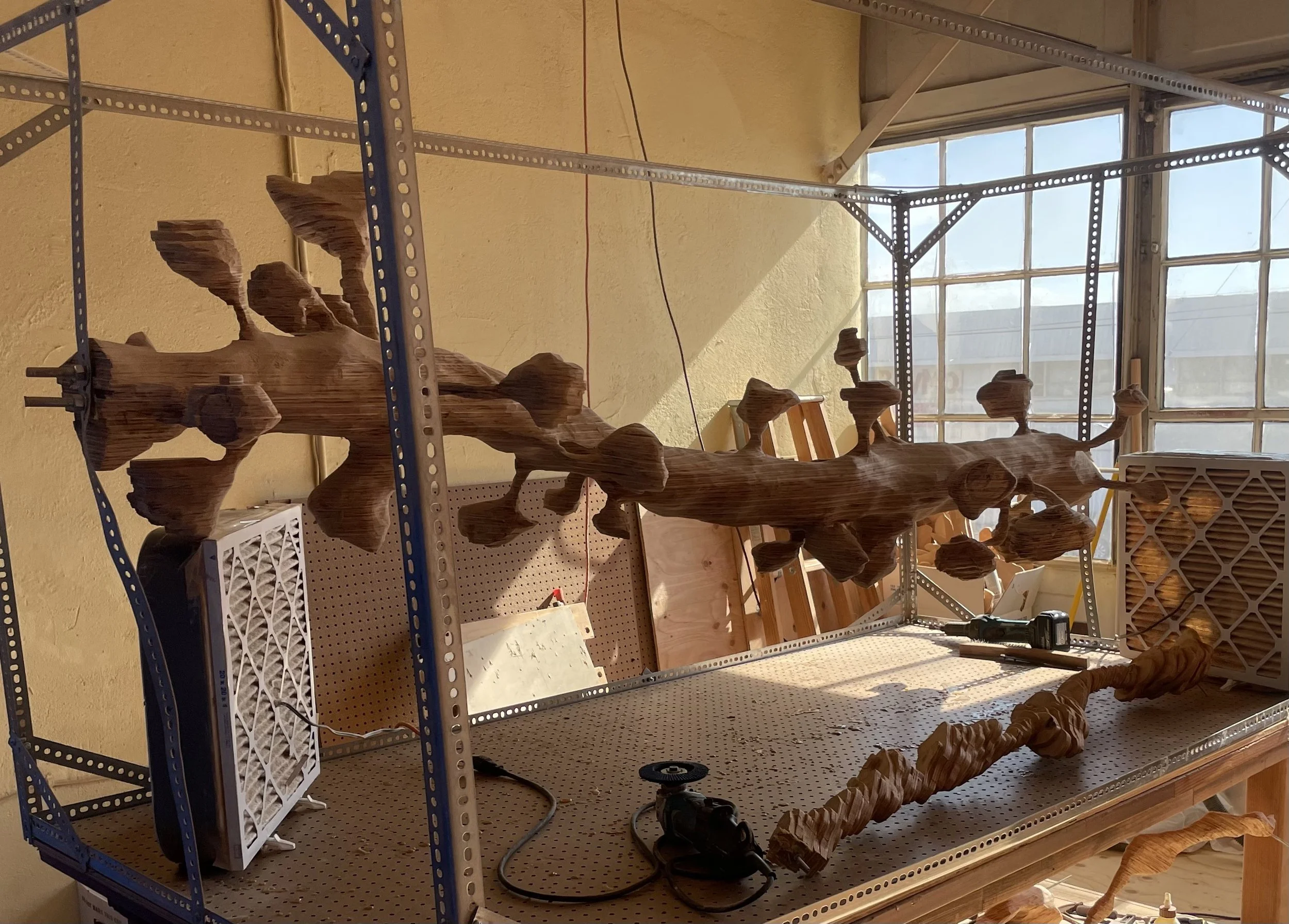
Mid stage sanding
Morning in the workshop
Chunky spines
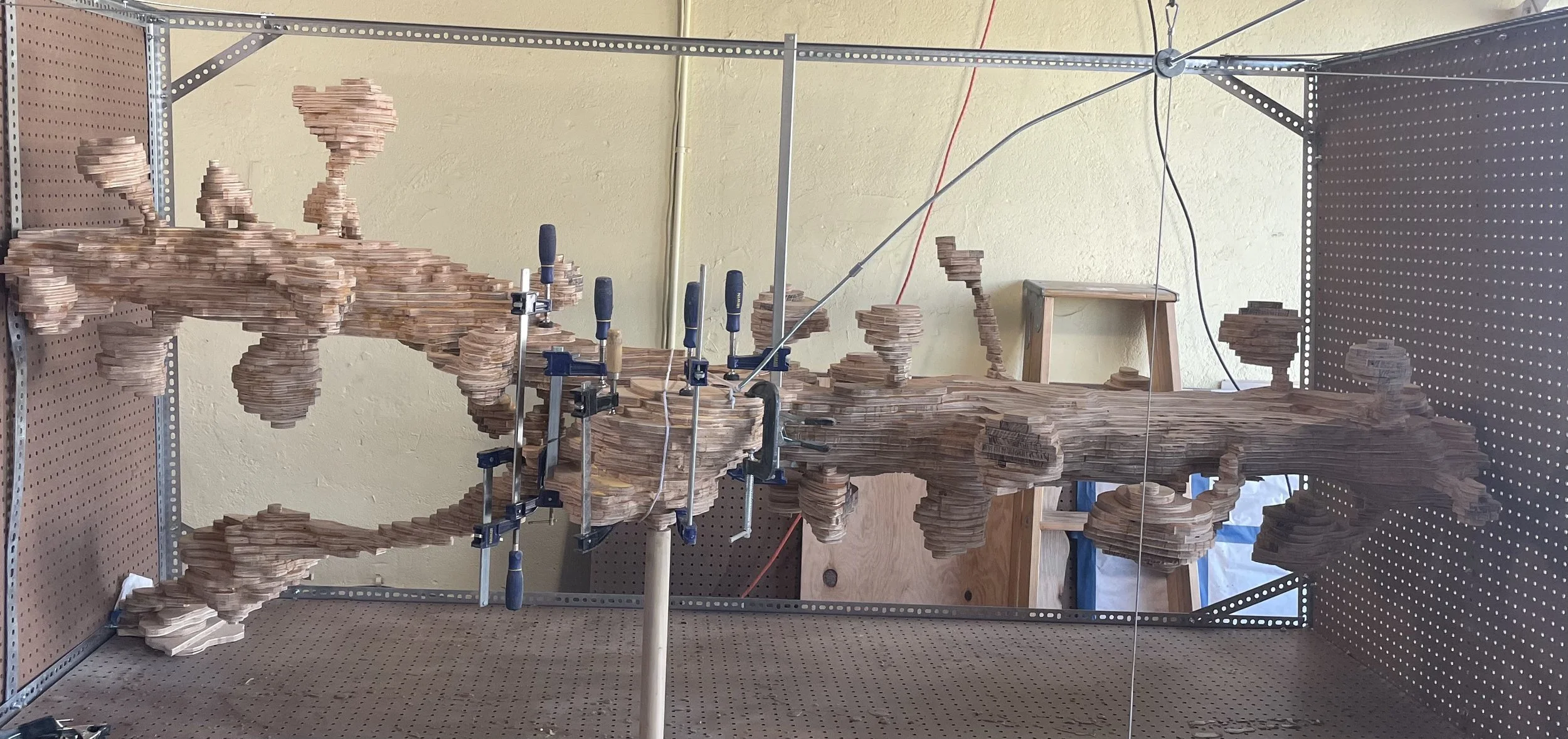
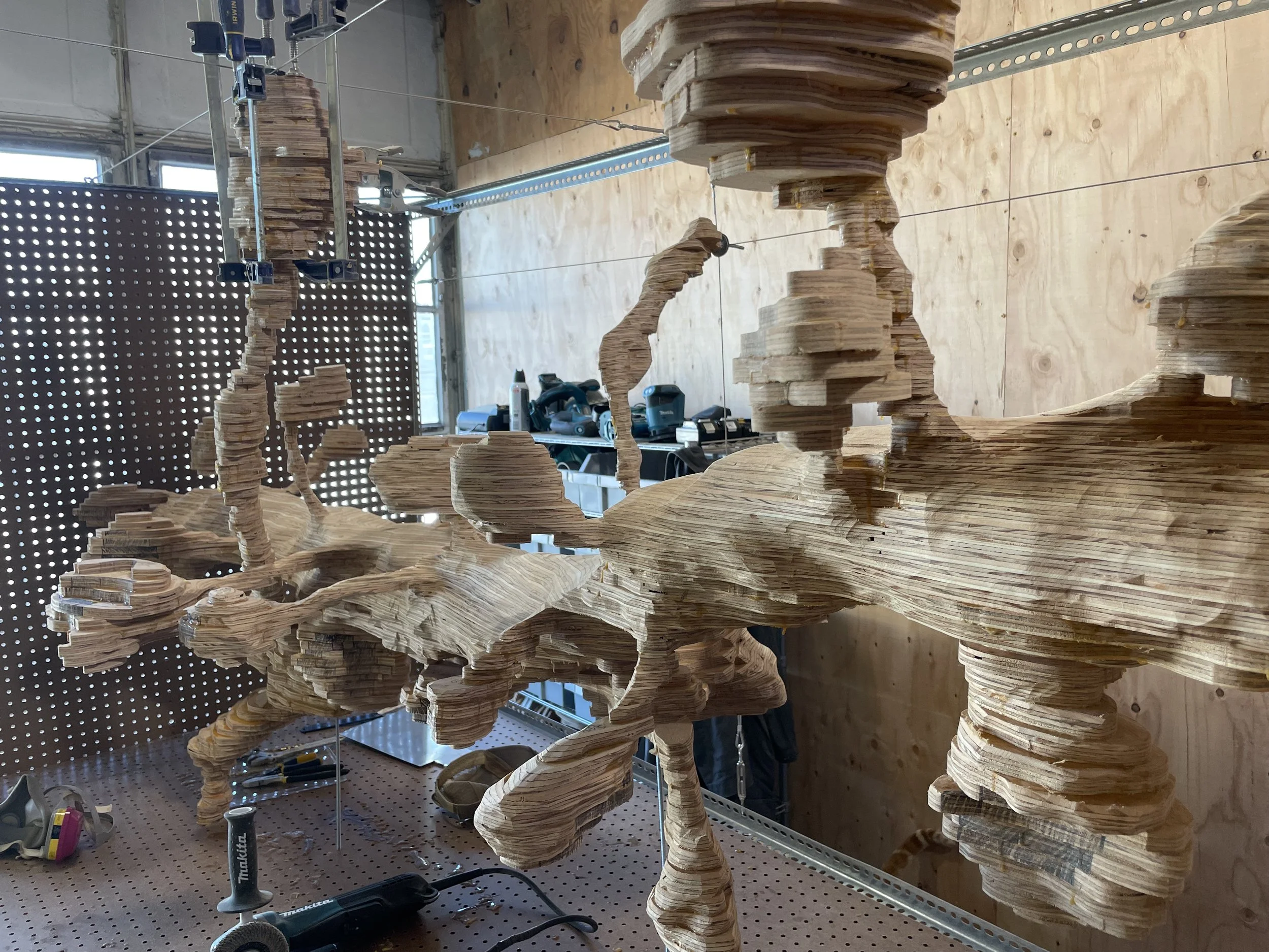
Adding inhibitory and excitatory axons
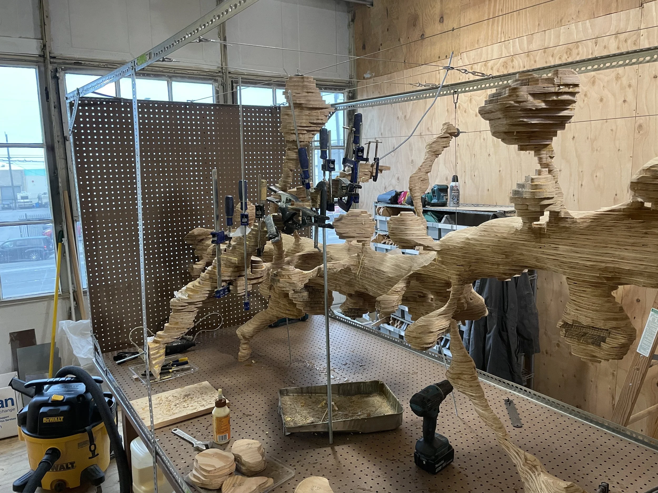
Building excitatory axons
Building excitatory axons
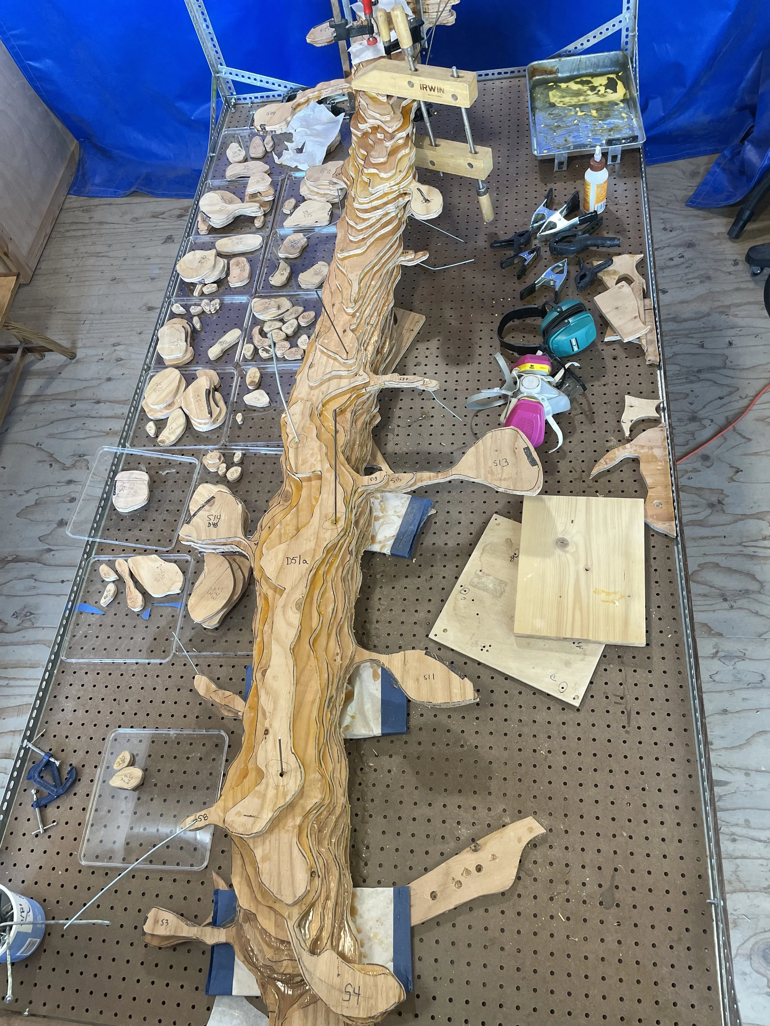
Laying out the spines for reconstruction
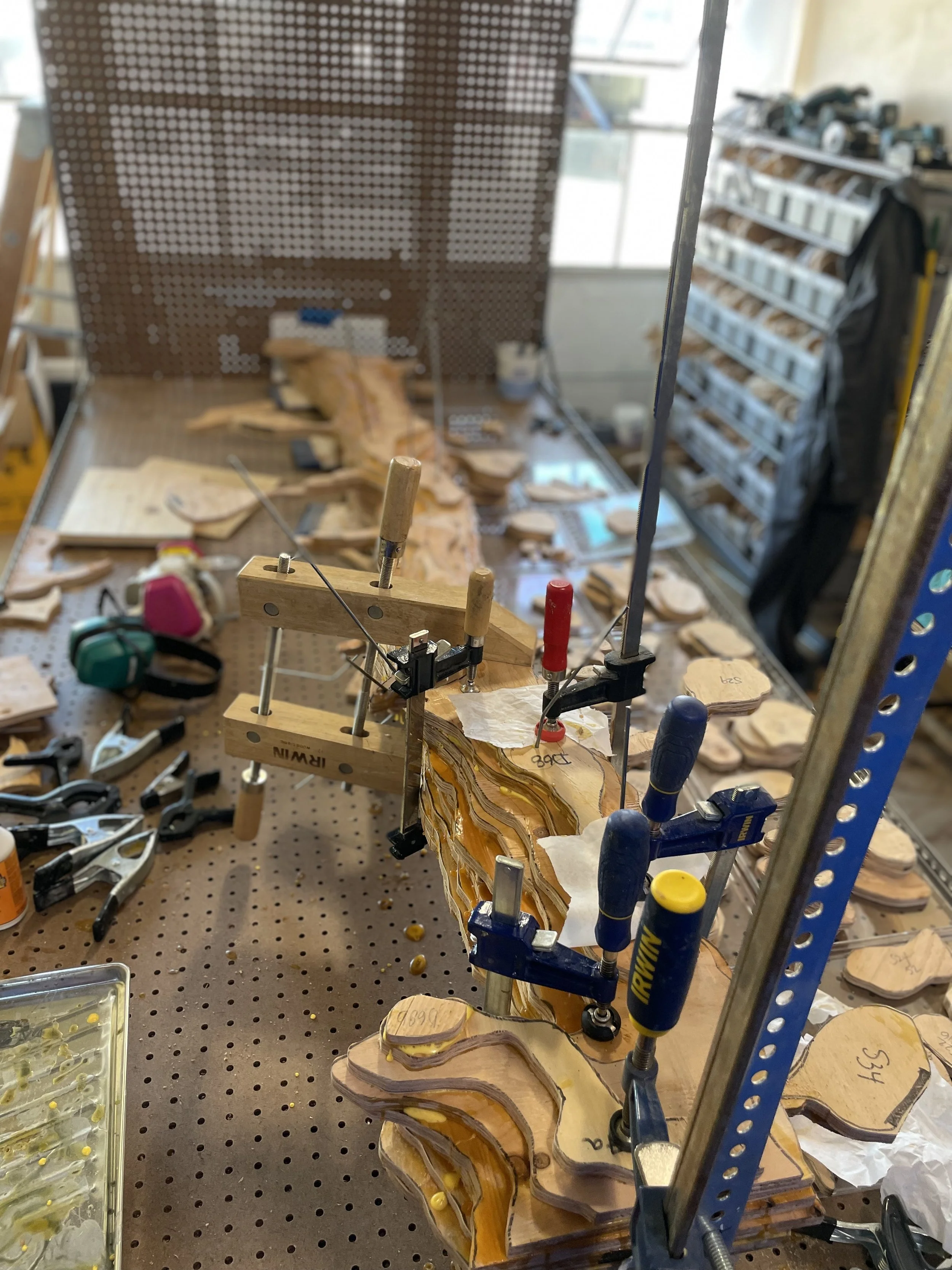
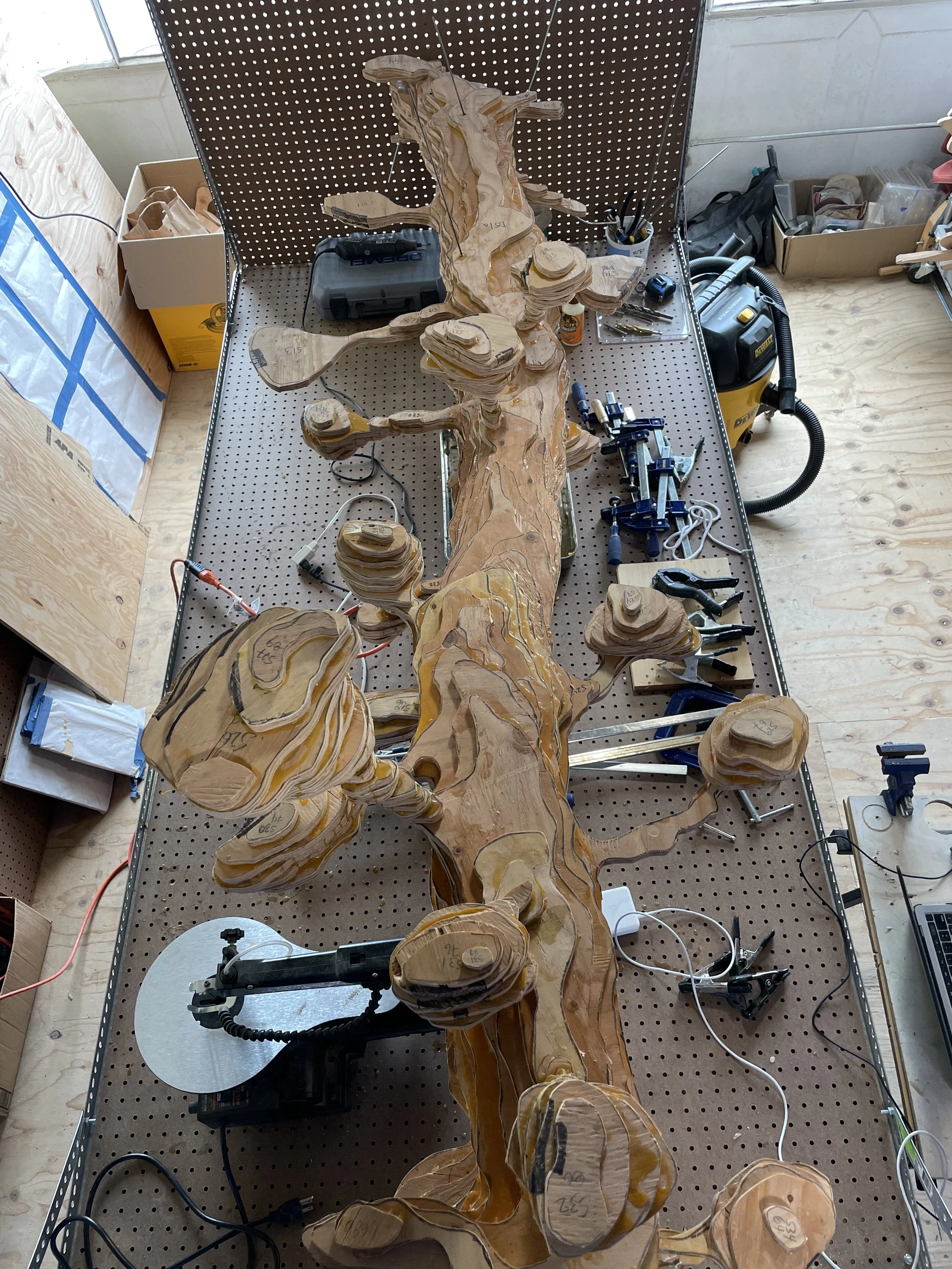
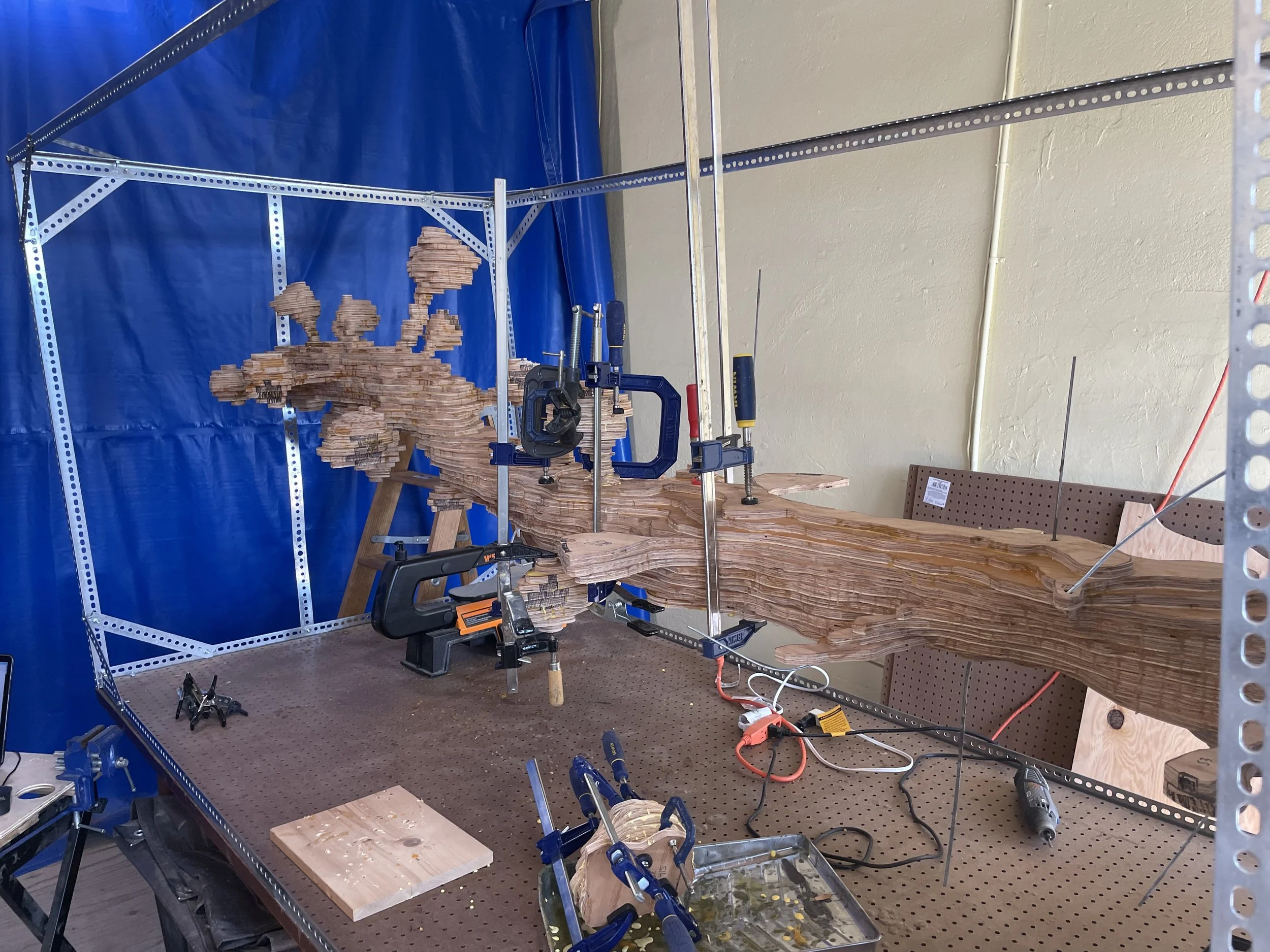
Gluing the core of the dendrite
Steel rod through the core of the dendrite
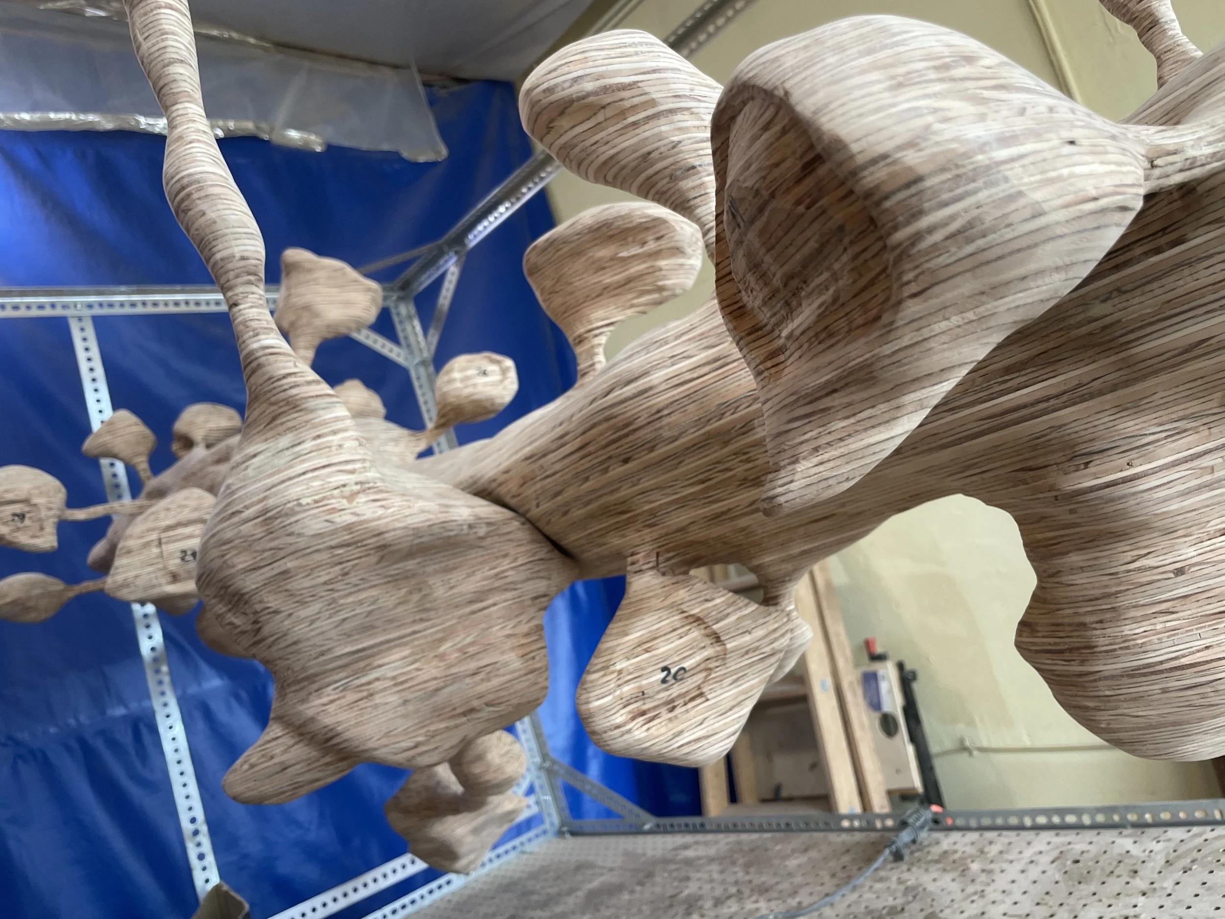
Early stage of dendrite reconstruction
First steps in dendrite reconstruction
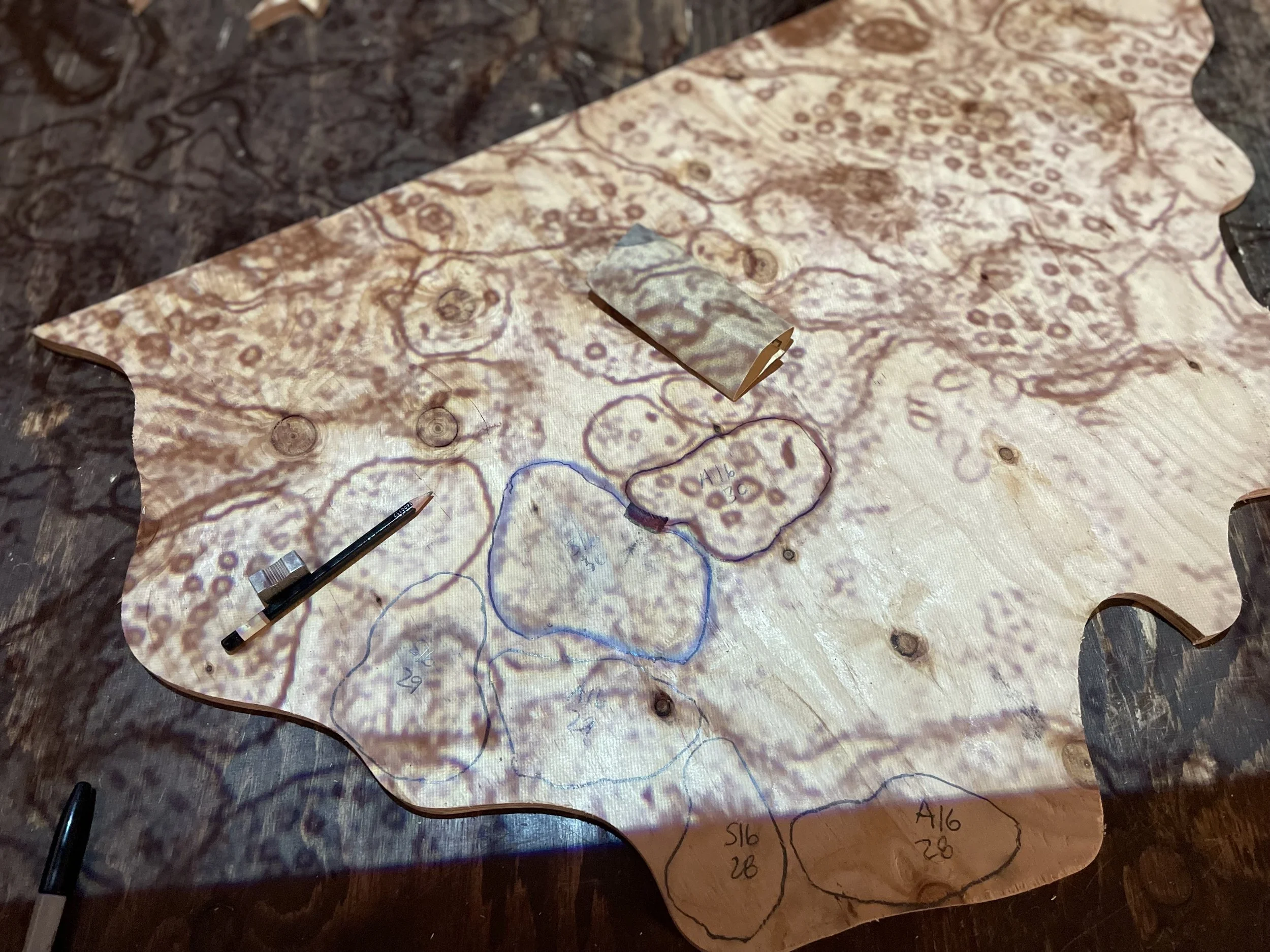
Mapping segmentation onto plywood
About the build
This is the biggest one I’ve done so far. It’s also the first using plywood. Using the plywood allowed me to go much bigger and more complex with the reconstruction. The even surface and rigidity of the plywood sheet is so different from thin cookies of redwood or pine I used in previous builds. Plywood is also an even and uniform thickness, so it circumvents a lot of the issues I ran into when cutting thin cookies with a chainsaw or even with a portable lumber mill. When I set out I didn’t fully appreciate that the plywood layering would generate such a neat topographic effect until I saw it emerge under the angle grinder. I think it turned out pretty well and I’m super pumped to do more with this.